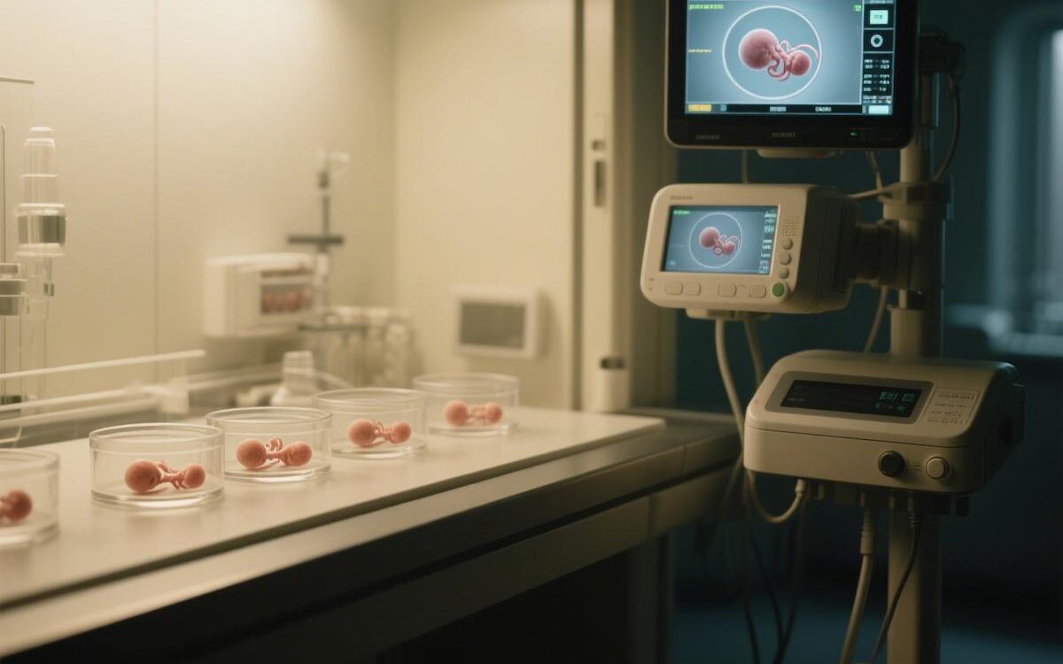
How does non-invasive technology select the “Chosen One” for IVF?
Each embryo is a unique code of life, which contains unlimited possibilities of future life. In the field of assisted reproduction, the core breakthrough in improving the success rate of IVF is the use of non-invasive IVF techniques to accurately select the “Chosen One” with the highest developmental potential, leaving behind the traditional era of “blind selection”. These techniques are just like installing a “see-through eye” for the embryo, deciphering its health code under the premise of zero damage, and lighting up the light of hope for countless families.
I. Embryos with Eyes of Fire: The Evolution of Morphology and Dynamics
Embryo evaluation has evolved from the initial static “face reading” to the dynamic tracking of the “dance of time”.
From static observation to dynamic tracking: Early reliance on static metrics such as prokaryotic patterns and cleavage sphere morphology was highly subjective. Time-lapse imaging has revolutionized the assessment method, like a “growth documentary” of the embryo, recording its developmental trajectory (fertilization cleavage, densification, blastocyst formation), and combining it with morphokinetic algorithms to provide a more objective and comprehensive prediction of the developmental potential.
AI-enabled precision “perspective”: AI deeply intervenes to analyze massive data (e.g., synchronization of cleavage, speed of blastocyst expansion) in time-lapse images. It can build virtual 3D models and even show strong predictive power at the chromosome level (e.g. aneuploidy identified with 83% accuracy). Research at the University of Cambridge Fertility Center has shown that AI-assisted screening significantly improves the efficiency of selecting high-quality embryos.
Advantages and unanswered questions: Morphological/kinetic assessment is suitable for very early embryos (e.g., single-cell stage), and time-delayed technology avoids the risk of embryo exposure and allows for more rapid assessment. However, its clinical superiority still needs to be confirmed by more large-scale randomized controlled trials (RCTs), and the cost of equipment investment is high.
II. Noninvasive decoding of DNA: a revolution in embryonic ploidy screening
Chromosome health (aneuploidy) is the cornerstone of successful embryo development. Aneuploidy is the main cause of fertilization failure and miscarriage.
Bottlenecks of traditional PGT-A: Preimplantation genetic testing (PGT-A) relies on trophoblast biopsy (invasive procedure), which has limitations such as damage to embryonic potential, inability to fully represent the chromosome status of the inner cell mass (developing fetus), and complexity of chimerism interpretation.
niPGT-A: The Rise of Liquid Biopsy: Non-Invasive Chromosome Screening (niPGT-A/NICS) is a disruptive breakthrough. It only requires analysis of free DNA (cfDNA) in embryo culture fluid, and allows precise assessment of embryo chromosome status through whole genome amplification (WGA) and high-throughput sequencing (e.g., NGS). The Johns Hopkins School of Medicine team demonstrated that its diagnostic accuracy is “on par” with traditional PGT-A, with excellent sensitivity and specificity.
Prospects and Challenges: niPGT-A is rapid, non-invasive, and has been successfully used for single embryo transfer screening (e.g., balanced translocation carriers). However, its widespread application still requires more RCTs to support a standardized process. In the future, the combination of AI morphologic analysis may provide a more efficient and cost-effective indirect assessment solution.
III.The “bug” of gene viability: transcriptomics
Early embryonic development relies on maternal reserves and subsequent activation of its own genome, whose activity is a key signal of potential.
RNA signaling: the “language” of the embryo: RNAs secreted by the embryo into the culture medium, especially non-coding RNAs (ncRNAs), are valuable biomarkers. MicroRNAs (miRNAs) and piRNAs have been shown to be deeply involved in gene regulation, preparation for implantation and maintenance of genomic stability.
Pinpointing the “code of success”: High-throughput sequencing technology identifies specific miRNAs (e.g. miR-199a-5p, miR-483-5p) and piRNAs that are strongly associated with embryo quality, and a study at Karolinska Institutet in Sweden utilized machine learning to reveal the presence of aneuploid blastocysts in successful pregnancies. Unique gene expression ‘fingerprints’.
A tool for rapid detection: Quantitative Real-Time PCR (qRT-PCR) allows for rapid, highly sensitive detection of these RNA markers, facilitating immediate clinical decision-making. However, it requires specialized personnel and equipment.
IV.The “Whisper” of Proteins: Signal Interpretation by Secretomics
Proteins secreted by the embryo are its language of communication with the outside world, and are a direct reflection of its developmental status and potential for implantation.
From simple assays to high-throughput analysis: Early days relied on ELISA to detect specific proteins (e.g. sHLA-G). Nowadays, mass spectrometry (e.g. MALDI-TOF MS) has become the mainstream technology, which can analyze massive protein profiles in cultures in a single, unbiased step.
Constructing predictive models: By comparing the secreted protein profiles of aneuploid/aneuploid or successful/failed embryos, researchers have built models with high predictive value. For example, a team at the University of Heidelberg, Germany, used MALDI-TOF MS to predict persistent pregnancies, showing excellent positive predictive values.
Overdrive and standardization challenges: The sample size required is extremely small (micro-escalation), the assay is extremely fast (can be performed instantly before transplantation), and the operation is relatively easy. However, the comparability of results across laboratories is affected by differences in media composition, industry standards are urgently needed, and the cost of equipment is an obstacle to diffusion.
V. Metabolic Barometer: Metabolomics Insights into Embryo Viability
The metabolic activity of embryos is a direct reflection of their energy status and developmental vigor, which is like a unique “metabolic fingerprint” of life.
Energy transitions and metabolic signatures: Embryonic development is accompanied by significant metabolic shifts (e.g., from pyruvate to glucose utilization). The consumption/accumulation pattern of nutrients (glucose, amino acids) in the culture medium and the content of specific metabolites (e.g. tyrosine, tryptophan) have been shown to be strongly correlated with the developmental potential of the embryo.
Spectroscopic “non-invasive probes”: techniques such as Raman and NIR can quickly “read” the metabolic fingerprints of culture fluids without touching the sample. A UCLA study combining Raman spectroscopy and machine learning demonstrated high specificity and sensitivity (71.5% accuracy) in predicting pregnancy.
Great potential, consensus to be achieved: Metabolomics technology is fast and user-friendly. However, differences in assays, sample processing, data analysis, and media types have led to a lack of standardized clinical protocols that have been widely adopted around the world.
VI.Microscopic messengers: the potential of extracellular vesicles (EVs)
Extracellular vesicles (EVs) are nanoscale “communication packages” secreted by the embryo, carrying DNA, RNA, proteins and other key information, and serving as a bridge between the embryo and the mother.
The EVs carry genomic DNA (gDNA), miRNAs, proteins, etc., which reflect the chromosomal status (ploidy), viability, and potential of the embryo for implantation. Studies at Massachusetts General Hospital (MGH) have shown that EVs secreted by successfully conceived embryos are unique in concentration, size and miRNA profile.
Multi-technology fusion assays: Nanoparticle tracking analysis (NTA) to determine the concentration and size of EVs; flow cytometry combined with specific markers to analyze their nucleic acid content; and genomics techniques (e.g., sequencing, aCGH) to decipher the genetic information they carry.
The future is bright, and validation is underway: EVs are a promising source of biomarkers because they are simple, rapid, relatively inexpensive, and completely non-invasive. However, their potential for clinical application is in dire need of a higher level of evidence-based support from large, rigorously designed RCTs.
VII. AI-enabled, Multidimensional Integration: The Intelligent Brain for Future Embryo Selection
It is difficult to depict the whole picture of embryo potential with a single indicator. Integrating multi-omics data with AI is an inevitable direction for building accurate prediction models.
AI: the “intelligent alchemist” of data: machine learning algorithms can efficiently integrate multidimensional data from morphokinetics, culture cfDNA, ncRNA, proteome, metabolome, EVs, etc., and even incorporate patients’ clinical information (age, medical history, and sperm parameters) to build powerful prediction models. The integrated model developed by the Stanford team significantly improved the identification of high-quality embryos.
Mapping a finer “blueprint of life”: Researchers are working to integrate data from single-cell sequencing and other sources to build a finer map of human embryonic development from fertilization to early organ formation, providing a deeper foundation for understanding potential.
The future is here: Although some early AI applications have been criticized as “publicity bubbles”, there is no doubt about their great potential to improve the objectivity, accuracy and efficiency of embryo screening. With the accumulation of data and optimization of algorithms, AI-driven non-invasive, multi-dimensional embryo evaluation will surely become the core engine of IVF success rate improvement.
VIII.Far-reaching impact: rewriting the trajectory of IVF families’ fortunate “pregnancy”.
The booming development of non-invasive embryo screening technology is profoundly reshaping the landscape of assisted reproduction:
For IVF families: significantly increase the success rate of IVF, reduce the physical and mental suffering and economic burden of repeated transfers; completely avoid the risk of biopsy damage; promote the popularization of single embryo transfer (SET), eliminate complications related to multiple pregnancies (preterm birth, low birth weight), and ensure the safety of mothers and infants; some technologies realize “same day evaluation – same day transfer”, and shorten the anxiety of waiting. Some of the techniques have realized “same-day evaluation – same-day transplantation”, thus shortening the anxiety of waiting.
For fertility doctors: provide objective and quantitative gold standard for embryo selection, optimize clinical decision-making; promote the standardization of embryo evaluation process; provide new weapons for difficult cases such as advanced age and repeated failures; reduce ethical dilemmas involving chimeric embryos.
For the industry chain: Drive innovation and market expansion of time-delay systems, sequencers, mass spectrometers, spectroscopy equipment and AI software; as the technology matures and scales up, the cost of testing is expected to drop and accessibility will be improved; accelerate the formation of industry technical standards and guidelines; and the massive amount of multi-homology data accumulated will provide invaluable insight for the research and development of new culture media and drugs.
Looking ahead
Non-invasive embryo screening technologies are iteratively converging at an unprecedented rate. Although new stars such as niPGT-A and EVs analysis still need to be validated by more large-scale RCTs, and the standardization of proteomics and metabolomics is in urgent need of a breakthrough, the direction of advancement is crystal clear: more accurate, safer, and more efficient.
Artificial Intelligence, as a “smart brain”, deeply integrates multi-omics “life big data” and draws a fine “potential map” of embryonic development, which will become the core key to unlock a higher IVF success rate. This will be the core key to unlocking a higher IVF success rate. Science is gently but firmly unveiling the initial veil of life, so that every precious embryo can run towards the end of blooming life under the most scientific guidance. Say goodbye to “blind selection” and embrace precision – this is the most precious gift of life from science and technology to future parents.