Clinical significance and systematic management guidelines for NT examination
I. Medical definition and technical principle of NT examination
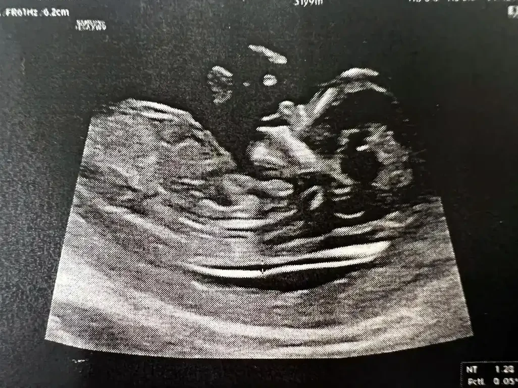
NT (nuchal translucency) refers to the transparent area formed by the accumulation of subcutaneous lymphatic fluid in the posterior part of the fetal neck, the maximum thickness of which is measured by high-frequency ultrasound (5-7 MHz probe) in the median sagittal section of the fetus. This structure is prominent at 11-14 weeks of gestation, as the fetal lymphatic system is not fully developed at this stage and some of the lymphatic fluid is temporarily retained. Chromosomal abnormalities (e.g., trisomy 21) can lead to structural abnormalities in the lymphatic vessels, which can obstruct the return of lymphatic fluid and increase the thickness of the NT. Studies show that 65% of fetuses with NT ≥3.5 mm have chromosomal abnormalities or severe congenital heart disease, and every 1 mm increase in NT raises the risk of trisomy 21 by 32 times.
Timing and operation standardization
According to the guidelines of the International Federation of Gynecology and Obstetrics (FIGO), NT examination should be strictly controlled from 11 weeks + 0 days to 13 weeks + 6 days (head and hip diameter 45-84 mm). Earlier than this, the NT is not sufficiently formed, and later than this, abnormal lymphatic fluid may be absorbed, leading to a missed diagnosis. There are three technical requirements to be met during the procedure:
The fetus must be placed in a natural supine position with the spine at a 45° angle to the probe;
The ultrasound image should clearly show the nasal bone, the fourth ventricle and the cleft palate;
The measurement point should be chosen at the widest part of the hyaloid layer, and the maximum value should be repeated three times with an error of <0.1 mm. Clinical grading and management pathway of the test results Low risk (NT <2.5 mm): probability of chromosomal abnormality <1%, routine mid-pregnancy ultrasound screening is recommended, with a focus on cardiac structures (e.g., venous catheterization flow spectrum); Critical risk (2.5-3.4 mm): combining maternal serum free β-hCG (median multiplicity MoM<0.5) and PAPP-A (MoM<0.4) increases the detection rate of trisomy 21 to 95%; if serologic abnormalities are present, noninvasive DNA testing (NIPT) is recommended rather than direct invasive procedures. Procedure; High risk (≥3.5 mm): referral to prenatal diagnostic center is required within 48 hours; chorionic villus sampling (CVS) is preferred to obtain placental tissues for rapid detection of chromosome aneuploidy of 13/18/21/X/Y by QF-PCR, and a preliminary report will be issued within 3 working days. IV. Refinement of special scenarios Multiple pregnancies: NT difference >20% between monochorionic twins suggests Twin-Twin Transfusion Syndrome (TTTS), amniotic fluid depth and umbilical artery flow need to be monitored every 2 weeks;
Structural anomalies combined with NT thickening: 40% risk of congenital heart disease if coexisting with nasal bone loss or tricuspid regurgitation, requiring fetal echocardiography at 16 weeks of gestation;
Normal karyotype but NT thickening: even if chromosomally normal, 15% of fetuses with NT ≥3.5 mm still have monogenic disorders such as Noonan syndrome, and whole exome sequencing (WES) is recommended.
V. Synergistic application with other screening tools
Early pregnancy combined screening (FCTS): NT combined with serum PAPP-A and β-hCG has a detection rate of 90% for Down syndrome and a false positive rate of 5%;
Noninvasive DNA testing (NIPT): for NT 2.5-3.4 mm pregnant women, NIPT can detect T21/T18/T13 with >99.7% sensitivity, but cannot assess structural malformations;
Serologic screening in mid-pregnancy: if early pregnancy screening is missed, the quadruple test (AFP+uE3+hCG+Inhibin A) at 15-20 weeks of gestation provides a 60%-70% detection rate, but needs to be combined with systematic ultrasound.
VI. Precision decision making for invasive diagnosis
Chorionic villus sampling (CVS): performed at 11-13 weeks of gestation, puncture needles enter the placental villi transabdominally or transcervically, 0.7% risk of miscarriage but placental-fetal karyotypic discordance (CPM phenomenon) may be encountered in 2% of cases;
Amniocentesis: performed after 16 weeks of gestation, karyotyping by amniotic fluid cell culture, 0.1%-0.3% risk of miscarriage, simultaneous detection of neural tube defects is possible (AFP>2.5 MoM suggests risk);
Complementary molecular techniques: chromosome microarray analysis (CMA) detects the presence of pathogenic copy number variants (CNVs) in 7% of NT-thickened fetuses, such as 22q11.2 microdeletion syndrome.
VII. Multidisciplinary collaboration for subsequent pregnancy management
Multidisciplinary consultation needs to be initiated after the diagnosis of an abnormal fetus:
Geneticists interpret CMA/WES results and assess the risk of recurrence;
Fetal medicine specialists to develop a plan for termination of pregnancy (before 24 weeks of gestation);
Neonatal surgery for correctable malformations such as congenital diaphragmatic hernia and planning the timing of surgery after birth;
Psychological support team to provide grief counseling to reduce the incidence of postpartum depression (40% reduction in depression scores in the intervention group).
VIII. Clinical application of emerging technologies
Artificial intelligence-assisted measurement: deep learning algorithms (e.g., AI-NT system) reduce operator dependency from 25% to 5%, with 95% measurement consistency;
Three-dimensional ultrasound flow imaging: assessing lymphatic vessel development by blood flow signals within the nuchal translucency improves the positive predictive value for Turner syndrome to 78%;
Exosomal biomarkers: maternal blood levels of PLAC4 mRNA were negatively correlated with NT thickness (r=-0.68) and can be used as a complementary risk assessment indicator.
The value of NT examination as a core component of early pregnancy screening lies not only in risk stratification, but also in providing a time window for subsequent precise intervention. In clinical practice, a standardized quality control system should be established, combined with rapid molecular diagnosis and multidisciplinary collaboration, so that 95% of serious fetal anomalies can be clearly diagnosed before 18 weeks’ gestation, and families can be provided with sufficient decision-making support and psychological buffer.